New Publication from Seda’s Joanna Denbigh on label-free imaging of complex drug delivery systems
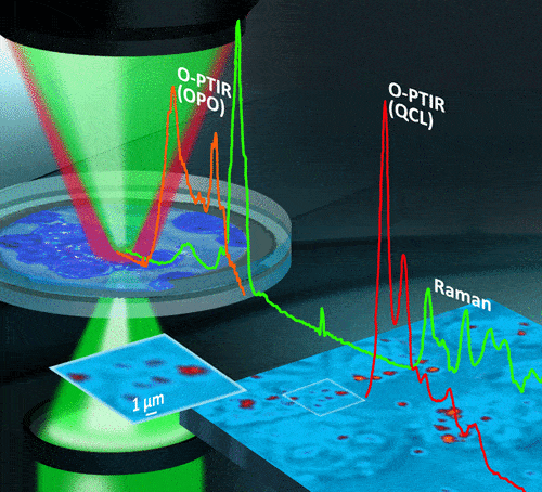
New paper on label-free imaging of complex drug delivery systems titled “Analysis of fixed and live single cells using optical photothermal infrared with concomitant Raman” has just been published in Analytical Chemistry.
Read the full publication here.
The authors describe a novel, high resolution, label-free imaging technique used successfully in live cells to analyse subcellular structures. This work represents a significant breakthrough for the spectroscopic analysis of biological systems, with broad applications including the characterisation of complex targeted drug delivery systems such as liposomes.
The paper reports the first use of a novel, completely optically based photothermal method (O-PTIR) for obtaining infrared spectra of both fixed and living cells using a quantum cascade laser (QCL) and optical parametric oscillator (OPO) laser as excitation sources, thus enabling all biologically relevant vibrations to be analysed at submicron spatial resolution. In addition, infrared data acquisition is combined with concomitant Raman spectra from exactly the same excitation location, meaning the full vibrational profile of the cell can be obtained. A major breakthrough was the ability to identify an undistorted amide I band in live cells in an aqueous environment, opening up the possibility of detecting subtle protein secondary structure changes, normally obscured by the presence of water.
IR spectral data are collected in a noncontact (far-field) mode and are obtained without any dispersive scatter artifact (such as the Mie scatter) that can plague conventional FTIR/QCL microscopes, thus improving the confidence of spectral interpretations of subtle spectral differences. The concomitant Raman spectra yield additional complementary information regarding the cells and demonstrate that this new combined methodology can provide enhanced spectroscopic analysis.
Specifically, the authors were able to:
- Identify organelles in fixed cells and live cells using both O-PTIR and Raman;
- Observe an increase in β-sheets or RNA bands, consistent with the nucleolus, which is the site where new ribosomes are assembled; therefore, it contains rRNA (essential component of the ribosomes) and proteins;
- Identify lipid-rich regions assigned to the endoplasmic reticulum from consideration of their localization in the cell (compared to our confocal fluorescence microscopy data) and the OPO IR image;
- Obtain, from the live cells in aqueous buffer, high-quality discrete frequency images despite the presence of the water layer, enabling the identification of lipid droplets ∼1 μm or less.

This work is the result of a collaboration between Seda Principal Scientist Dr Joanna Denbigh, and eminent academics from the University of Manchester including Professor Jayne Lawrence (NowCADD Director) and Professor Peter Gardner. The first author of this paper is Dr Alice Spadea, a NoWCADD funded postdoctoral researcher and the work has been carried out in partnership with Mustafa Kansiz, Director of Product Management and Marketing at Photothermal Spectroscopy Corp.
Work was supported by the North West Centre for Advanced Drug Delivery (NoWCADD), a collaborative partnership between the Division of Pharmacy and Optometry, University of Manchester, and AstraZeneca.
